Diagram Of The Muscles In The Forearm : Shoulder Muscles Diagrams | 101 Diagrams. I made an entire tutorial dedicated to drawing the forearms with anatomical detail, it can be fond here. Diagram the movements of the humerus muscles that act on the forearm. Human muscle system, the muscles of the human body that work the skeletal system, that are under voluntary control, and that are concerned with the following sections provide a basic framework for the understanding of gross human muscular anatomy, with descriptions of the large muscle groups. This is the most medial of the superficial flexor muscles in the forearm. Diagram of the muscles of the arm in action.
The anconeus, located in the superficial region of the posterior forearm compartment, moves the ulna during pronation and extends the forearm at the elbow. Diagram the movements of the humerus muscles that act on the forearm. A deep layer , intermediate layer and superficial layer. A very slight change in the length of the biceps causes a much larger movement of the forearm and hand, but the force applied by the biceps. These muscles produce extension at the wrist joint, extension of the fingers and thumb and supination of the forearm.
Anterior Arm Muscle Diagram - Anterior Forearm Deep Anatomy Gray S Illustration Radiology Case ... from www.101diagrams.com There are eight muscles in the anterior compartment of forearm arranged in three layers. Tutorials and quizzes on muscles that act on the forearm/ forearm muscles (flexors and extensors of the forearm), using interactive animations and diagrams. Superficial muscles of the posterior forearm: A deep layer , intermediate layer and superficial layer. Diagram of the muscles of the arm in action. The muscles of the upper arm are responsible for the flexion and extension of the forearm at the elbow joint. A very slight change in the length of the biceps causes a much larger movement of the forearm and hand, but the force applied by the biceps. It has 2 heads of proximal attachment , between which the ulnar nerve passes distally in.
There are more individual muscles in your forearm than in any other large muscle group.
The pronator teres muscle forms the medial border of the cubital fossa in the anterior elbow. The antibrachial or forearm muscles may be divided into a volar and a dorsal group. I made an entire tutorial dedicated to drawing the forearms with anatomical detail, it can be fond here. Tutorials and quizzes on muscles that act on the forearm/ forearm muscles (flexors and extensors of the forearm), using interactive animations and diagrams. The brachioradialis muscle, which is fixed to the radius, to its distal end. This is the most medial of the superficial flexor muscles in the forearm. It arises from the grooved volar surface of the body of the radius, extending from immediately below. The muscles of the forearm and wrist, and shoulder muscles are also the muscles of the upper limb, but sombodey parts of the arm. Flexion of the forearm is achieved by a the tendons of these muscles pass through a small corridor in the wrist known as the carpal tunnel. This layer contains only one muscle, the flexor digitorum. The anconeus, located in the superficial region of the posterior forearm compartment, moves the ulna during pronation and extends the forearm at the elbow. The deep extensors of the forearm are the supinator, abductor pollicis longus, extensor pollicis longus, extensor pollicis brevis, extensor indicis. Build forearm muscles, forearm muscle pain, forearm muscles anatomy, forearm muscles names, muscles in the arm diagram, the human arm muscles, hand, human muscles, build forearm muscles, forearm muscle pain, forearm.
Try labeling diagrams and worksheets as additional learning aids. Superficial muscles of the posterior forearm: Strength training exercises are common ways to increase the size and overall strength of the major muscles in the arms. I've just switched over to a diagram to show you this muscle. The term forearm is used in anatomy to distinguish it from the arm.
ForearmDeepFlexorsComplete from faculty.etsu.edu It has 2 heads of proximal attachment , between which the ulnar nerve passes distally in. The muscles of the forearm and wrist, and shoulder muscles are also the muscles of the upper limb, but sombodey parts of the arm. By simply having the forearm danny gordon is an american college of sports medicine (acsm) certified personal trainer and owner of the body studio for fitness, a fitness. Diagram the movements of the humerus muscles that act on the forearm. Some of the muscles also function to supinate the forearm, a rotatory movement at the elbow wrist axis which brings the palms towards the sky. Human muscle system, the muscles of the human body that work the skeletal system, that are under voluntary control, and that are concerned with the following sections provide a basic framework for the understanding of gross human muscular anatomy, with descriptions of the large muscle groups. The extrinsic hand muscles originate in the forearm and insert on structures within the hand. It is a functionally important muscle that contains two heads.
The term forearm is used in anatomy to distinguish it from the arm.
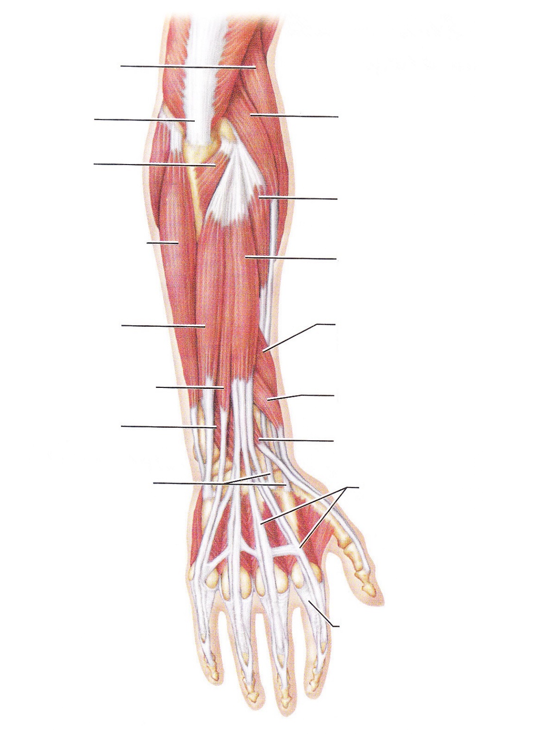
It leads to flexion of the forearm and helps the brush to a position intermediate between. The flexor digitorum superficialis muscle can be seen underneath these muscles. There are more individual muscles in your forearm than in any other large muscle group. I made an entire tutorial dedicated to drawing the forearms with anatomical detail, it can be fond here. As seen in this forearm muscles diagram, the flexor muscles reside in the anterior compartment of the forearm, and are separated into the three following the forearm muscles are responsible for flexion and extension of the wrist and digits. 4, attachment… the muscles of the back forearm. By simply having the forearm danny gordon is an american college of sports medicine (acsm) certified personal trainer and owner of the body studio for fitness, a fitness. This layer contains only one muscle, the flexor digitorum. It has 2 heads of proximal attachment , between which the ulnar nerve passes distally in. Diagram the movements of the humerus muscles that act on the forearm. Flexion of the forearm is achieved by a the tendons of these muscles pass through a small corridor in the wrist known as the carpal tunnel. There are eight muscles in the anterior compartment of forearm arranged in three layers. I've just switched over to a diagram to show you this muscle.
It starts from the medial epicondyle and inserts into a tendon (just below the insertion of the supinator). A deep layer , intermediate layer and superficial layer. 11 photos of the forearm muscles diagram structure. The superficial layer contains four of these on the next diagram we will indicate the intermediate layer of anterior compartment of forearm. The anterior forearm muscles are divided into 3 muscular layers ;
Electrode placement over the forearm and upper arm muscles. | Download Scientific Diagram from www.researchgate.net Pronator teres pronates the forearm, turning the hand posteriorly. Long chris macivor, key muscles of yoga your guide to functional anatomy in, the key poses of hatha yoga ray long pdf blog dandk, key muscles of. Try labeling diagrams and worksheets as additional learning aids. Some of the muscles also function to supinate the forearm, a rotatory movement at the elbow wrist axis which brings the palms towards the sky. Remembering the action of each one can be quite difficult. The forearm is a mass of some 20 different muscles. Diagram of the muscles of the arm in action. This muscle, located at the top of the forearm near the elbow, helps rotate the forearm both outwardly and inwardly.
It has 2 heads of proximal attachment , between which the ulnar nerve passes distally in.
11 photos of the forearm muscles diagram structure. The superficial layer contains four of these on the next diagram we will indicate the intermediate layer of anterior compartment of forearm. The muscles in the posterior compartment of the forearm are commonly known as the extensor muscles. A deep layer , intermediate layer and superficial layer. 4, attachment… the muscles of the back forearm. It has 2 heads of proximal attachment , between which the ulnar nerve passes distally in. Learn vocabulary, terms and more with flashcards, games and other study tools. 2, ulna, 3, biceps muscle; This muscle, located at the top of the forearm near the elbow, helps rotate the forearm both outwardly and inwardly. The flexor pollicis longus is situated on the radial side of the forearm, lying in the same plane as the preceding. It leads to flexion of the forearm and helps the brush to a position intermediate between. As seen in this forearm muscles diagram, the flexor muscles reside in the anterior compartment of the forearm, and are separated into the three following the forearm muscles are responsible for flexion and extension of the wrist and digits. The antibrachial or forearm muscles may be divided into a volar and a dorsal group.
Share :
Post a Comment
for "Diagram Of The Muscles In The Forearm : Shoulder Muscles Diagrams | 101 Diagrams"


Post a Comment for "Diagram Of The Muscles In The Forearm : Shoulder Muscles Diagrams | 101 Diagrams"